What is Aplastic paleness?
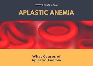
Aplastic iron deficiency happens when the bone marrow delivers excessively not many of each of the three sorts of platelets: red platelets, white platelets, and platelets. A decreased number of red platelets cause hemoglobin to drop. A diminished number of white platelets make the patient vulnerable to disease. Furthermore, a decreased number of platelets cause the blood not to cluster as without any problem.
What causes Aplastic sickliness?
Aplastic pallor has different causes. A portion of these causes are idiopathic, which means they happen inconsistently for no known explanation. Different causes are optional, coming about because of a past ailment or confusion. Gained causes, be that as it may, may incorporate the accompanying:
1. History of explicit irresistible illnesses like irresistible hepatitis
2. History of taking certain medications, such as antibiotics and anticonvulsants
3. Exposure to specific poisons like weighty metals
4. Exposure to radiation
5. History of an immune system illness
What are the manifestations of aplastic paleness?
Coming up next are the most widely recognized indications of aplastic iron deficiency. Be that as it may, every individual may encounter manifestations in an unexpected way.
How is aplastic paleness analyzed?
Notwithstanding a total clinical history and actual assessment, symptomatic techniques for frailty incorporate extra blood tests and a bone marrow biopsy.
Treatment and steady treatment may include:
1. Blood bonding (both red platelets and platelets)
2. Excessive anti-infection treatment
3. Meticulous hand washing
4. Special consideration to food arrangement, (for example, just eating cooked food sources)
5. Avoiding building locales which might be a wellspring of specific organisms
6. Medications (to animate the bone marrow to deliver cells)
7. Immunosuppressive treatment and chemical treatment
Hemolytic weakness
Weakness coming about because of a diminished red cell life expectancy. In extreme cases, red cell life expectancy might be a couple of days.
Grouping of Hemolytic weakness
I. Hereditary:
Film deserts:
Genetic spherocytosis Hereditary elliptocytosis
Metabolic:
G6PD inadequacy PK insufficiency
Haemoglobinopathy:
eg Sickle (HbS), Hemoglobin C (Hb)
II. Acquired:
Safe interceded:
Auto antibodies
Warm immunizer Cold counter acting agent
Alloantibodies
Bonding related hemolysis Hemolytic sickness of the infant
III. Drugs and Chemicals:
Red cell Fragmentation:
Contamination causing hemolysis Malaria
Clostridia
Paroxysmal Nocturnal Hemoglobinuria (PNH)
General clinical highlights of Hemolytic anemias:
Whiteness, gentle jaundice, splenomegaly, folate insufficiency (overabundance use) prompting megaloblastic iron deficiency and to aplastic emergencies.
Research facility Findings
Pallor
Raised unconjugated bilirubin Low serum haptoglobins
Reticulocytes Spherocytosis on blood film
In the event that the hemolysis is intravascular: hemoglobin anemia, hemoglobinuria, haemosiderinuria, methane albuminuria
Inherited Spherocytosis
Autosomal prevailing. Deformity in the particular segment of the red cell film which brings about a difference in red cell shape to a spherocytic. Phorocytes get caught in the spleen where resulting glucose hardship improves breakdown.
Clinical and Laboratory Features Presents at whatever stage in life Fluctuating jaundice Gallstones
Reticulocytotic Spherocytic
Research facility tests
Osmotic delicacy expanded
Auto hemolysis expanded (rectified by glucose) Coombs test negative
Treatment
Splenectomy – (be careful irresistible confusions a while later, so immunize with Pneumovax and hemophilic flu first).
Inherited elliptocytosis
Like HS, yet red cells are circular. Typically a milder problem.
Red Cell protein abandons
1. G6PD insufficiency: Several hereditary varieties. Normal in West Africa, Mediterranean, Middle East and South East Asia. Anyway can happen anyplace on the planet.
Set off by oxidant stress
1. Drugs
2. Fava beans
3. Infections
Clinical Features: Those of intravascular hemolysis, with hemoglobinuria and iron deficiency.
Determination:
During an emergency – blood film shows ‘nibble cells’ (Heinz bodies eliminated by the spleen). Heinz bodies are oxidized, thick hemoglobin particles in the red cell.
Formal determination is by estimation of G6PD compound in cells (immediate and circuitous tests).
Treatment: Remove encouraging oxidant factor.
2. Pyruvate Kinase lack: Rare Autosomal latent Blood film shows mutilated cells (poikilocytes) as ‘prickle cells’.
Gained Hemolytic anemias
Hemolysis can be intravascular (bringing about the arrival of free hemoglobin) or extra vascular (by take-up of red cells into the reticuloendothelial framework).
Invulnerable
(Immune system haemolytic anaemias – AIHA)
Because of creation of immunizer coordinated against red cell antigens. The direct antoglobulin test (Coombs) is positive. Autoantibodies respond at various temperatures, bringing about the terms ‘warm’ and ‘cold’ immune system haemolytic frailty.
Warm AIHA
Ordinarily IgG (with or without noticeable supplement restricting – C3d). At times IgA or IgM.
Cells become spherocytes, and are taken up overwhelmingly in the spleen, and the reticuloendothelial framework.
Etiology
Related with Systemic Lupus Erythematosis (SLE), Chronic Lymphocytic Leukemia (CLL), or medication prompted or idiopathic.
Medication incited AIHA
Neutralizer can be coordinated against the medication and red cell, or immunizer against medication can fix supplement on the red cell surface.
Clinical Features
Paleness, Splenomegaly
Frequently an ongoing backsliding and dispatching course can happen along with ITP (‘Evans condition’)
Research center discoveries
Spherocytosis, positive direct antiglobulin (Direct Coombs) test at 37& degree.
Treatment
First Line: Steroids Resistant cases: a few choices
– High portion intravenous immunoglobulin
– Cyclosporine
– Splenectomy
Patients are regularly folic corrosive lacking as a result of high red cell turnover – so supplements should be given.
Cold AIHA
Neutralizer connects to red cells in the cold for example at limits, or when blood is taken into a venesection bottle. This element is especially connected with IgM antibodies.
Etiology
Mycoplasma pneumonia Infectious mononucleosis
Paraoxsyomal Cold Haemoglobinuria (PCH) – generally found in youngsters after a viral contamination Lymphoma
The immunizer is generally coordinated against explicit red cell antigens for example I or I, however in PCH, it is coordinated against P (the Donath Land Steiner immune response).
Clinical highlights
A persistent Hemolytic frailty aggravated by cold. Pallor, jaundice, splenomegaly, purple skin staining (acrocyanosis) at limits
Post disease types (Mycoplasma, irresistible mononucleosis) are normally transient.
Research center highlights
Spherocytosis (less set apart than warm AIHA)
Positive DAGT (typically uncovering C3 supplement just on a superficial level without counter acting agent discovery).
Treatment
Keep patient warm
Immunosuppressive treatment for example steroids, cyclophosphamide or chlorambucil
© 2021 Niazi TV – Education, News & Entertainment


