A neurotransmitter is a structure in the unmistakable system that starts a neuron (or nerve cell) to move an electrical or compound picture to another neuron or to the efferent target cell.
Presentation
The term neurotransmitter is gotten from syn indicating “together” and haptenic implying “closing.” The substance or electrical sign from one nerve cell (neuron) to another nerve cell is moved by a structure called neural association. In that limit, in neuronal correspondence, neurotransmitters help. Synaptic structures are changed by synaptic bond particles (SAMs) that are set up.
Structure
Neurons are not in direct contact with each other, they are detached by neurotransmitters known as little openings. The fundamental portion of the neurotransmitter is the advancement of movement possibilities between neurons. The scope of drugs limits by interfering with synapses related with synaptic transmission.
The Structure of The Neurotransmitter
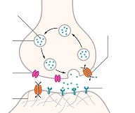
An enormous segment of the intersection focuses between neurons appear as neurotransmitters. Portions of axons lay near dendrites of various neurons yet don’t interface; between them there is a void of around 20um. This void is crossed by the arrival of a synapse from the axon film (pre-synaptic layer), which diffuses over the space to breath life into the dendritic film (post-synaptic layer), at the stage where points of view are sent.
The Constraint of the Neurotransmitter is To Pass on Activity Possibilities Between Neurons.
• Data correspondence between neurons
• Synapses arise as unions where nerve motivations move one heading explicitly (from the pre-synaptic neuron to the post-synaptic neuron): considering the way that the synaptic vesicles are simply present in the presynaptic neuron bulb and the synapse receptors are expressly described in the post-synaptic film
• Filter out low level upgrades. That is, discard ‘foundation change’ from the material structure. If insufficient synapse is passed on from the pre-synaptic bulb into the synaptic isolated (by then inadequate synapse will tie to the receptors on the post-synaptic layer and lacking Na+ atom channels will open to permit the film potential to get to – 55mV). These low level redesigns don’t cause nerve primary purposes to advance to the post-synaptic neuron.
• To shield the reaction structure from over-provoking
The presence of a development potential at the pre-synaptic handle (pre-synaptic bulb) reinforces voltage gated Calcium (Ca2+) atom channels to open, permitting Ca2+ particles to enter the pre-synaptic bulb. The inundation of calcium particles causes the synaptic vesicles to converge with the pre-synaptic film so passing on the synapse (for example acetylcholine) into the synaptic segregated.
Precisely when the synapse diffuses over the opening (synaptic isolated), it partners with a receptor site on the post-synaptic layer, depolarizing it thusly starting an activity potential and nerve motivation in the going with neuron. The postsynaptic layer contains unequivocal protein receptors with which the neural association particles join. Exactly when joined, Na+ atom organizes open in the film, permitting Na+ particles to diffuse from the split into the postsynaptic neuron.
In the event that the film winds up being adequately depolarized, a development potential is started in the axon of the postsynaptic neurone (an activity potential will conveyed at the “axon hillock” where the cell body contacts plot the axon, if enough Na+ particles diffuse to this point from the cell body to depolarize the layer potential to – 55mV cutoff: all the Na+ atom channels as of now will by then open permitting smart flood of Na+ particles into the axon and making an activity potential = ALL OR NOTHING law).
Acetylcholine, when passed on, is immediately beat by proteins in the synaptic confined, so its impact is restricted and the converging of principle driving forces is thwarted. In the event that lacking acetylcholine is passed on, the postsynaptic film won’t be engaged. The compound which beats acetylcholine is called cholinesterase. The following choline and ethanoic ruinous diffuse back over the synaptic split to change acetylcholine inside the pre-synaptic bulb. ATP is required to re-structure transmitter particles and store them in vesicles. Another transmitter substance is noradrenaline. It happens, close by acetylcholine, in the modified material structure.
Electrical or Substance Transmission?
Close to the completion of the nineteenth and beginning of the 20th century, there was a huge load of discussion about whether synaptic transmission was electrical or compound.
• Some people accepted that motioning over a synapse incorporated the movement of particles authentically from one neuron into another—electrical transmission.
• Other people thought it depended upon the appearance of an engineered from one neuron, causing a response in the getting neuron—substance transmission.
Compound transmission is more ordinary, and more bewildered, than electrical transmission. Thusly, we ought to explore compound transmission first.
Graph of Transmission at Synthetic Neurotransmitters
Manufactured transmission incorporates appearance of substance dispatches known as neural connections. Neurotransmitters pass on information from the pre-synaptic—sending—neuron to the post-synaptic—getting—cell.
An action expected goes down the axon of the presynaptic—sending—cell and appears at different axon terminals veering from the axon. The axon terminal is close by the dendrite of the postsynaptic—tolerating—cell. This spot of close relationship among axon and dendrite is the synapse.
A single axon can have various branches, allowing it to make neural associations on various postsynaptic cells. Basically, a single neuron can get countless synaptic commitments from a wide scope of presynaptic—sending—neurons.
Inside the axon terminal of a sending cell are various synaptic vesicles. These are film bound circles stacked up with neural connection particles. There is a little opening between the axon terminal of the presynaptic neuron and the layer of the postsynaptic cell, and this opening is known as the synaptic separated.
Picture demonstrating pre-synaptic cell’s axon terminal containing synaptic vesicles with neurotransmitters. Voltage-gated calcium channels are apparently surface of the axon terminal. Over the synaptic split, there is the post-synaptic cell surface solicited in receptors (ligand-gated molecule channels) for the neurotransmitter.
Exactly when an action potential, or nerve drive, appears at the axon terminal, it institutes voltage-gated calcium coordinates in the cell film. Ca2+text{Ca}^{2+}Ca2+C, a, start superscript, at least 2, end superscript, which is accessible at significantly higher concentration outside the neuron than inside, hustles into the cell. The Ca2+text{Ca}^{2+}Ca2+C, a, start superscript, at least 2, end superscript licenses synaptic vesicles to merge with the axon terminal layer, conveying neurotransmitter into the synaptic isolated.
Picture demonstrating what happens when action possible appears at axon terminal, causing molecule stream and depolarization of target cell. Step by step:
1. Action potential shows up at axon terminal and depolarizes layer.
2. Voltage-gated calcium channels open and calcium particles stream in.
3. Calcium molecule storm triggers synaptic vesicles to convey neurotransmitter.
4. Neurotransmitter binds to receptors on target cell (for this circumstance, making positive particles stream in).
The particles of neurotransmitter diffuse over the synaptic isolated and bind to receptor proteins on the postsynaptic cell. Authorization of postsynaptic receptors prompts the opening or closing of molecule coordinates in the cell film. This may be depolarizing—make inside the cell more certain—or hyperpolarizing—make inside the cell more negative—dependent upon the particles being referred to.
Now and again, these effects on channel direct are prompt: the receptor is a ligand-gated molecule channel, as in the diagram above. In various cases, the receptor isn’t a molecule channel itself yet orders molecule channels through a hailing pathway. See the article on neurotransmitters and receptors for more information.
