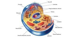
The inside of the cell is separated into the core and the cytoplasm. The core is a round or oval-formed design at the focal point of the cell. The cytoplasm is the area outside the core that contains cell organelles and cytosol, or cytoplasmic arrangement. Intracellular liquid is all things considered the cytosol and the liquid inside the organelles and core.
Layers
Layers are the doors to the cell. The plasma film is the specific obstruction encompassing the cell. It gives an obstruction to the development of atoms between the intra-and extracellular liquids. Review that extracellular methods outside the cell. The plasma film additionally serves to moor nearby cells together and to the extracellular network. Different signals and sources of info can change the affectability and penetrability of layers.
The Fluid Mosaic Model: film structure
Films are made of a twofold layer of lipids, primarily phospholipids, containing inserted proteins. The inserted proteins are significant as facilitators in moving particles through the layer. The actual film is coordinated into a bimolecular layer, implying that the non-polar district is coordinated in the center (away from water as it is hydrophobic) and the polar areas are situated toward the outside: the extracellular liquid and the cytosol. Another approach to consider it is two lines of pins with their heads to the outside and the needle part to within. Heads, needles, needles, heads. Like a sandwich. As the phospholipid particles are not artificially bound to one another and hence every atom is allowed to move freely, the general bi-layer structure has an adaptable smoothness. Cholesterol atoms are additionally installed in the plasma layer and serve to convey substances to cell organelles by shaping vesicles.
The proteins installed in the layer are ordered into two classes.
Fringe film proteins will be proteins on the layer surface, principally the cytosolic side where they communicate with cytoskeletal components to impact cell shape and motility. These proteins are not amphipathic and are bound to polar areas of the vital proteins.
Vital film proteins length the whole width of the layer, in this way getting through both the polar and non-polar districts of the design. These proteins can’t be eliminated from the layer without upsetting the lipid bilayer.
Understand that layer capacities are subject to the compound organization and any deviations in arrangement between the two surfaces of the film and the particular proteins that are joined to or related with the layer. The plasma film additionally has an extracellular surface layer of monosaccharides that are connected to the layer lipids and proteins. This layer is known as the glycocalyx and is significant in the intercellular acknowledgment measure.
Film Junctions
Integrins are transmembrane proteins that tight spot to explicit proteins in the extracellular lattice and to film proteins on nearby cells. Integrins help to put together cells into tissues. They are additionally answerable for sending signals from the extracellular framework to the phone inside.
On the off chance that two cells are adjoining, yet isolated, they might be junctured by desmosomes. Desmosomes are thick collections of protein at the cytoplasmic surface of the plasma films of both separate cells. They are invaded with protein strands that reached out into one or the other cell. The reason and capacity of desmosomes is to hold neighboring cells solidly set up in regions that are liable to extending, like skin.
Another sort of film intersection is the tight intersection. These intersections are framed by the real actual joining of the extracellular surfaces of two neighboring plasma films. Tight intersections are significant in regions where more power over tissue measures is required, for example, the epithelial cells in the digestive system that are engaged with ingestion.
At long last, hole intersections are real protein channels that connect the cytosols of nearby cells. The downside to this ‘immediate connection’ is that it just allows more modest atoms to go through.
Cell Organelles
Cell organelles are the little workhouses inside the cell. Every one of the elements of life happen in every individual cell. Organelles can be delivered by breaking the plasma layer, through homogenization and ultracentrifuging the blend. The organelles are of various size and thickness and will settle out at explicit rates.
The core is in the focal point of most cells. A few cells contain various cores, like skeletal muscle, while some don’t have any, like red platelets. The core is the biggest film bound organelle. In particular, it is answerable for putting away and communicating hereditary data. The core is encircled by a specific atomic envelope. The atomic envelope is made out of two films joined at standard stretches to shape roundabout openings called atomic pores. The pores permit RNA particles and proteins adjusting DNA articulation to travel through the pores and into the cytosol. The choice cycle is constrained by an energy-subordinate interaction that changes the distance across of the pores because of signs. Inside the core, DNA and proteins partner to frame an organization of strings called chromatin. The chromatin gets imperative at the hour of cell division as it turns out to be firmly dense along these lines shaping the rodlike chromosomes with the enmeshed DNA. Inside the core is a filamentous area called the nucleolus. This fills in as a site where the RNA and protein parts of ribosomes are gathered. The nucleolus isn’t layer bound, yet rather a district.
Ribosomes are the destinations where protein atoms are orchestrated from amino acids. They are made out of proteins and RNA. A few ribosomes are discovered bound to granular endoplasmic reticulum, while others are free in the cytoplasm. The proteins incorporated on ribosomes bound to granular endoplasmic reticulum are moved from the lumen (open space inside endoplasmic reticulum) to the Golgi device for discharge outside the cell or circulation to different organelles. The proteins that are blended of free ribosomes are delivered into the cytosol.
The endoplasmic reticulum (ER) is aggregately an organization of layers encasing a particular constant space. As referenced before, granular endoplasmic reticulum is related with ribosomes (giving the outside surface a harsh, or granular appearance). Some of the time granular endoplasmic reticulum is alluded to as unpleasant ER. The granular ER is associated with bundling proteins for the Golgi mechanical assembly. The agranular, or smooth, ER needs ribosomes and is the site of lipid blend. What’s more, the agranular ER stores and deliveries calcium particles Ca2+.
The Golgi device is a membranous sac that serves to change and sort proteins into secretory/transport vesicles. The vesicles are then conveyed to other cell organelles and the plasma film. Most cells have at any rate one Golgi device, albeit some may have various. The device is typically situated close to the core.
Endosomes are layer bound rounded and vesicular constructions situated between the plasma film and the Golgi device. They serve to sort and direct vesicular traffic by squeezing off vesicles or combining with them.
Mitochondria are probably the main designs in the human body. They are the site of different compound cycles engaged with the blend of energy bundles called ATP (adenosine triphosphate). Every mitochondrion is encircled by two films. The external film is smooth, while the internal one is collapsed into tubule structures called cristae. Mitochondria are extraordinary in that they contain limited quantities of DNA containing the qualities for the blend of some mitochondrial proteins. The DNA is acquired exclusively from the mother. Cells with more noteworthy action have more mitochondria, while those that are less dynamic have less requirement for energy-creating mitochondria.
Lysosomes are limited by a solitary film and contain exceptionally acidic liquid. The liquid goes about as processing compounds for separating microbes and cell trash. They assume a significant part in the cells of the resistant framework.
Peroxisomes are likewise limited by a solitary film. They devour oxygen and work to drive responses that eliminate hydrogen from different atoms as hydrogen peroxide. They are significant in keeping up the substance adjusts inside the cell.
The cytoskeleton is a filamentous organization of proteins that are related with the cycles that keep up and change cell shape and produce cell developments. The cytoskeleton additionally frames tracks along which cell organelles move moved by contractile proteins connected to their different surfaces. Like a little parkway framework inside the cell. Three kinds of fibers make up the cytoskeleton.
Microfilaments are the most slender and generally plentiful of the cytoskeleton proteins. They are made out of actin, a contractile protein, and can be amassed and dismantled rapidly as indicated by the necessities of the cell or organelle structure.
Halfway fibers are marginally bigger in measurement and are discovered most widely in areas of cells that will be exposed to pressure. Desmosomes in the skin will contain fibers. When these fibers are amassed they are not equipped for quick dismantling.
Microtubules are empty cylinders made out of a protein called tubulin. They are the thickest and generally unbending of the fibers. Microtubules are available in the axons and long dendrite projections of nerve cells. They are fit for quick get together and dismantling as per need. Microtubules are organized around a phone area called the centrosome, which encompasses two centrioles made out of 9 arrangements of intertwined microtubules. These are significant in cell division when the centrosome creates the microtubular shaft filaments fundamental for chromosome partition.
© 2021 Niazi TV – Education, News & Entertainment
