Protozoan Parasites
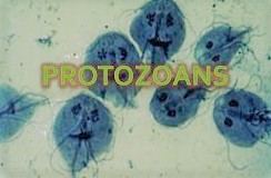
Protozoan contaminations are parasitic sicknesses brought about by living beings once in the past grouped in the Kingdom Protozoa. They incorporate living beings arranged in Amoebozoa, Excavate, and Chromalveolata. Models incorporate Entamoeba histolytica, Plasmodium (some of which cause intestinal sickness), and Giardia lamblia. Trypanosoma brucei, sent by the tsetse fly and the reason for African dozing ailment, is another model. They are treated with antiprotozoal specialists.
Here are the most widely recognized protozoan parasites in people.
•Balantidium Coli is a protozoan parasite living in the gastrointestinal lot. It causes an illness called balantidiasis.
•Entamoeba histolytica – Amoebiasis
•Giardia intestinalis
•Leishmania is a tiny parasite in the circulation system.
•Plasmodium vivax/falciparum – causes jungle fever (an extreme blood illness) in people.
•Toxoplasma gondii is an intracellular protozoa that causes toxoplasmosis.
•Trypanosoma brucei causes African Trypanosomiasis, the resting infection.
Amoebic Parasites
•Amoebiasis, otherwise called amebiasis or entamobiasis, is a contamination brought about by any of the single adaptable cells of the Entamoeba bunch. Manifestations are generally basic upon contamination by Entamoeba histolytica. Amoebiasis can give no, gentle, or serious manifestations. Indications may incorporate stomach torment, gentle the runs, bleeding loose bowels or serious colitis with tissue passing and hole. This last inconvenience may cause peritonitis. Individuals influenced may create iron deficiency because of loss of blood.
• nvasion of the intestinal coating causes amoebic wicked diarhea or amoebic colitis. In the event that the parasite arrives at the circulation system it can spread through the body, most regularly winding up in the liver where it causes amoebic liver abscesses. Liver abscesses can happen without past loose bowels. Growths of entamoeba can get by for as long as a month in soil or for as long as 45 minutes under fingernails. It is imperative to separate among amoebiasis and bacterial colitis. The favored indicative technique it through fecal assessment under magnifying lens, yet requires a talented microscopist and may not be dependable while barring disease. Expanded white platelet include is available in extreme cases, however not in gentle ones. The most precise test is for antibodies in the blood, yet it might stay positive after treatment.
•Prevention of amoebiasis is by isolating food and water from dung and by legitimate sterilization measures. There is no antibody. There are two treatment choices relying upon the area of the contamination. Amoebiasis in tissues is treated with one or the other metronidazole, tinidazole, nitazoxanide, dehydroemetine or chloroquine, while luminal disease is treated with diloxanide furoate or iodoquinoline. For treatment to be compelling against all phases of the single adaptable cell may require a blend of prescriptions. Diseases without manifestations don’t need treament yet contaminated people can spread the parasite to other people and treatment can be thought of. Treatment of other entamoeba contaminations separated from E. histolytica isn’t required.
•Infections can at times keep going for quite a long time. Side effects take from a couple of days to half a month to create and show themselves, yet ordinarily it is around two to about a month. Side effects can go from gentle looseness of the bowels to extreme diarrhea with blood and bodily fluid. The blood comes from sores framed by the amoebae attacking the covering of the digestive organ. In about 10% of obtrusive cases the amoebae enter the circulation system and may go to different organs in the body.
•It doesn’t as a rule interact with the digestive tract itself because of the defensive layer of bodily fluid that lines the gut. Illness happens when one-celled critter interacts with the cells coating the digestive tract. It at that point secretes similar substances it uses to process microbes, which incorporate catalysts that annihilate cell layers and proteins. This cycle can prompt infiltration and assimilation of human tissues, coming about first in flagon molded ulcers in the digestive system. Entamoeba histolytica ingests the obliterated cells by phagocytosis and is frequently seen with red platelets (an interaction known as erythrophagocytosis) inside when seen in feces tests.
Cause
Amoebiasis is a disease brought about by the single adaptable cell Entamoeba histolytica Likewise amoebiasis is some of the time erroneously used to allude to contamination with other amoebae, yet carefully talking it ought to be held for Entamoeba histolytica contamination. Other amoebae contaminating people include:
Parasites
• Dientamoeba fragilis, which causes Dientamoebiasis
• Entamoeba dispar
• Entamoeba hartmanni
• Entamoeba coli
• Entamoeba moshkovskii
• Endolimax nana and
• Iodamoeba butschlii.
Aside from Dientamoeba, the parasites above are not idea to cause infection.
Amoebiasis is normally communicated by the fecal-oral course, yet it can likewise be sent by implication through contact with grimy hands or articles just as by butt-centric oral contact. Disease is spread through ingestion of the pimple type of the parasite, a semi-torpid and solid design found in excrement. Any non-encysted amoebae, or trophozoites, pass on rapidly in the wake of leaving the body yet may likewise be available in stool: these are once in a while the wellspring of new diseases.
Analysis:
•With colonoscopy it is feasible to recognize little ulcers of between 3–5mm, yet determination might be troublesome as the mucous layer between these regions can look either solid or excited.
•Asymptomatic human contaminations are normally analyzed by discovering sores shed in the stool. Different buoyancy or sedimentation systems have been created to recuperate the pimples from fecal matter and stains help to picture the disconnected growths for tiny assessment.
•Since pimples are not shed continually, at least three stools ought to be inspected. In suggestive diseases, the motile structure (the trophozoite) can frequently be seen in new defecation. Serological tests exist and most people (if with manifestations) will test positive for the presence of antibodies.
Treatment: E. histolytica diseases happen in both the digestive system and in tissue of the digestive tract as well as liver. Subsequently, two distinct classes of medications are expected to treat the contamination, one for every area. Such enemy of amoebic medications are known as amebicides.
Control: To help forestall the spread of amoebiasis around the home:
•Wash hands altogether with cleanser and hot running water for at any rate 10 seconds in the wake of utilizing the latrine or changing a child’s diaper, and prior to taking care of food.
•Clean restrooms and latrines regularly; give specific consideration to latrine seats and taps.
•Avoid sharing towels or face washers.
•Avoid crude vegetables when in endemic zones, as they may have been treated utilizing human excrement.
•Boil water or treat with iodine tablets.
•Avoid eating road food sources particularly openly puts where others are sharing sauces in a single holder
•Good sterile practice, just as mindful sewage removal or treatment, are essential for the counteraction of E. histolytica contamination on an endemic level.
•E. histolytica growths are generally impervious to chlorination, in this manner sedimentation and filtration of water supplies are important to decrease the frequency of contamination.
© 2021 Niazi TV – Education, News & Entertainment
