Osmosis
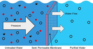
Assimilation is the net dispersion of water across a layer. Aquaporins are proteins that structure directs in the lipid bi-layer for the polar water atoms to diffuse through. There will be a net dissemination of the two compartments prompting dispersion balance with no adjustment of volume in one or the other compartment if the compartments are isolated by a film that is porous to both a solute and water. Nonetheless, if the film is simply penetrable to water (for example not to the solute) then, at that point dispersion balance will be reached with a net expansion in the volume of the compartment that had a higher osmolarity, regardless. Osmolarity is the absolute solute convergence of an answer and is estimated in units called osmosis.
Thusly, water focus in an answer is adversely related with the quantity of solute particles. Osmotic pressing factor is the pressing factor that should be applied to forestall the net progression of water into an answer isolated by a film. The osmotic pressing factor increments with expansions in osmolarity. Water will then, at that point move from locales of lower osmotic strain to districts of higher osmotic pressing factor.
At the point when a framework arrives at balance, the osmolarities of intra-and extracellular liquids are something very similar. An isotonic arrangement is an answer wherein cells will neither swell nor shrivel, this is expecting that the cells are set into an answer of non-infiltrating solutes with a similar osmolarity as the extracellular liquid. Interestingly, there is no net development in an isotonic arrangement. In a hypotonic arrangement, the arrangement contains less non-entering solutes, and the phones, along these lines, ingest water and the phones swell. At last, a hypertonic arrangement is one in which the arrangement contains more non-infiltrating solutes and water moves out of the cells and they contract. It is imperative to comprehend that infiltrating solutes don’t add to the constitution of the arrangement.
Endocytosis
Endocytosis is a transportation interaction that requires energy. The fundamental system is that areas of the plasma film crease into the cell which shapes little pockets within the cell. These pockets squeeze off into film bound vesicles inside the cell.
Liquid endocytosis alludes to when the vesicles framed to encase a little volume of extracellular liquid. Notwithstanding, if certain particles in the extracellular liquid end up restricting to explicit proteins on the plasma layer and are then conveyed into the phones with extracellular liquid, the interaction is then called adsorptive endocytosis. By and large, these two cycles are additionally called pinocytosis and are exhibited by most cells.
A few cells will inundate huge unfamiliar particles by means of an interaction called phagocytosis. This just occurs in particular cells that are generally very few and event. The sort of particles inundated incorporate microorganisms and cell garbage.
Endosomes are normally melded with endocytic vesicles sooner or later simultaneously, and the substance of the bundles are then passed into organelles like Lysosomes.
Both pinocytosis and phagocytosis are instances of endocytic measures. The large thing to recall is that the development of particles is from an external perspective of the plasma film to within.
Exocytosis
To move things from within the cell to the outside, film bound vesicles in the cytoplasm will intertwine with the plasma layer and delivery their substance outside the cell. The bound vesicle material then, at that point acclimatizes into the plasma layer. In this style, segments of the plasma film lost during endocytosis can be supplanted. Furthermore, the interaction gives a course by which film impermeable atoms, like protein chemicals, that are incorporated by cells can be delivered into the extracellular liquid. At last, the interaction of exocytosis is set off by improvements that lead to an increment in cytosolic calcium fixation which thus enacts proteins needed for the vesicle film to meld with the plasma layer and consequently fixing any ‘openings’ from earlier cycles.
Epithelial Transport
The luminal (or apical or mucosal) layer is the plasma film surface of an epithelial cell that faces an empty or liquid filled chamber. The basolateral (or serosal) film is the outside of the plasma layer on the contrary side generally neighboring an organization of veins.
Substances can cross a layer of epithelial cells through two pathways:
The paracellular pathway alludes to dispersion between nearby cells in the epithelium. This pathway is restricted to little particles and water due to the presence of tight intersections.
The transcellular pathway alludes to the development into an epithelial cell from one side, then, at that point dissemination through the cytosol and exit through the contradicting layer.
The vehicle and penetrability qualities of the luminal and basolateral films are not the equivalent because of the presence of various particle channels and carriers. Substances, in this manner, can move from a locale of lower fixation on one side to a higher focus on the other.
Glands
Organ cells discharge natural atoms integrated by their own cell cycles and they additionally emit salts and water, moving them starting with one extracellular compartment then onto the next.
The pace of discharge is constrained by substance or neural signals and work by:
Adjusting the pace of union
Adjusting the pace of exocytosis by means of calcium channels
Adjusting the siphoning pace of carriers and opening pace of particle channels
Two kinds of Glands:
Endocrine organs will deliver their emissions straightforwardly into the interstitial liquid encompassing the organ cells.
Endocrine organs emit chemicals.
Exocrine organs use ventilation work to associate with epithelial surfaces. The emissions move through the ventilation work or onto the outside of the epithelium. Sweat and salivary organs are instances of exocrine capacity.
© 2021 Niazi TV – Education, News & Entertainment
