Introduction
The primary individual actually to contemplate and portray cells was Robert Hooke in 1665, who inspected the little gaps under perhaps the most punctual magnifying lens that made up a cut of stopper he was watching. Those cell relics were dead and void so he had no clue about what a living cell’s inward structure was like.
The first time Anton van Leeuwenhoek recognized living cells (unicellular protists) under a magnifying lens was not until 1674. He named them animalcules (“little creatures”) since they moved under their own control, not understanding that they were just single cells. The advancement of cell hypothesis was trailed by further perceptions and work by Theodor Schwann, Matthias Jakob Schleiden, and Rudolf Virchow.
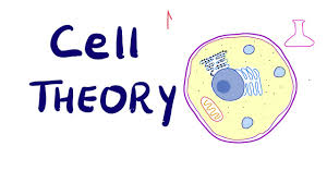
I. Cell Theory
The first cell hypothesis expressed that
• All living beings are made out of at least one cells.
• The cell is the fundamental auxiliary and utilitarian unit of life.
• Cells emerge from previous cells by a cycle of cell division. Present day revelations have included that
• Energy stream happens inside cells by means of digestion and biochemical responses.
• Genetic/heritable data is passed from cell to cell upon division.
• All cells have a similar fundamental compound sythesis.
Evaluate that there are two specific types of cells, Prokaryotic and Eukaryotic. Microscopic species and archaeans are prokaryotes (from the Greek master, meaning “previously” and karyon, meaning “nut” which makes no reference to the dim recoloring core prokaryotes). Any other species is eukaryotic, like protists, creatures, people, and plants.
Allude to your talk notes and text readings to audit the essential parts and life systems of prokaryotic and eukaryotic cells. Prokaryotic DNA is carried on a solitary, round chromosome, while eukaryotic DNA is sorted out on different, straight chromosomes that go through a composed duplication and isolation known as mitosis. Albeit prokaryotic separation in two (by means of splitting) to recreate, they don’t go through a mitotic cell cycle like that portrayed here, which is one of a kind to eukaryotes.
II. The Cell Cycle
Without understanding the structure and potential of a living cell, one can’t genuinely understand science. Proliferation is one of the most essential capacities of cells, regardless of whether agamic (mitosis) or sexual (meiosis). As it isolates, the cell goes through (to some degree subjectively characterised) stages.
On the off chance that you watch a populace of developing cells, regardless of whether part of a life form’s body or in a culture dish, you are probably going to see cells in different periods of the cell cycle . The phases of dynamic mitosis have been named for simple referral to occasions happening in each stage.
A. Interphase
Cells invest most of their energy in interphase. An interphase cell is caught up with going through the synthetic responses that encourage vitality move, developing, and getting ready to separate.
Interphase isn’t important for mitosis, which is characterized as dynamic cell division. Or maybe, it is the living phase of the cell. The chromosomes can’t be seen effectively, as they are in the diffuse structure known as chromatin. The qualities on the chromosomes are effectively being interpreted and deciphered, with the specific qualities being communicated relying upon the personality and capacity of the specific cell. The stages known as G1, S, and G2 happen during interphase, and are engaged with ordinary cell development guideline (G1), DNA blend (S), and protein and microtubule combination going before mitosis (G2).
B. Prophase
Prophase is the main phase of mitosis. The chromosomes consolidate and get obvious. Despite the fact that they have just copied, the chromosomes are not yet noticeable as isolated sister chromatids. The atomic film separates, and the shaft strands start to shape at inverse posts of the cell.
C. Metaphase
In this second period of mitosis, the copied chromosomes (presently called sister chromatids) loosen up from one another, and are joined distinctly at a somewhat tightened district, the centromere.
It is just since the chromosomes take on the “X” shape so regularly found in outlines. This “X” really speaks to not one, yet two chromosomes: the indistinguishable sister chromatids delivered during DNA replication.
Axle strands append to the kinetochore, a protein structure situated at the centromere, and the chromosomes are organized along the equator of the cell.
D. Anaphase
Axle strands abbreviate, the kinetochores discrete, and the chromatids (presently little girl chromosomes) are pulled separated and start moving to inverse shafts.
