The tissue on which evocator works and the tissue reactions is known as responsive tissue. The activity of the pointer through evocator is known as enlistment activity or coordinator activity. This cycle of enlistment impacts incredibly the protein union component of responsive tissues because of which unequivocal structure shaping cells become dynamic.
Starting Point of The Idea of The Coordinator
Spemann’s test (1924): A German embryologist Hans Spemann and his understudy Hilde Mangold (1924) performed transplantation investigate a newt Triturus cristatus, a Urodela of class Amphibia. Spemann united a piece taken from the dorsal lip of early gastrula of Rana sp. to the horizontal lip locale of the early gastrula of Triturus cristatus.
The undeveloped organism of Rana sp. is contributor and the incipient organism of Triturus is the host. They saw that the cells of the united piece go into the gastrula and structure notochord and somites. In this incipient organism its own dorsal lip of blastopore structures neural section, notochord, mesoderm and so forth
Also the united tissue impacts to shape notochord, neural notch and mesoderm. That is in a similar undeveloped organism twofold arrangement of notochord, nerve line and mesoderm are delivered. For this situation giver tissue has emitted some synthetic substances which has instigated to shape neural depression, notochord and so on in the host undeveloped organism.
The giver tissue had shades and the prompted neural section has additionally hued colors. After the fulfillment of the gastrulation they saw that a hatchling has created with two heads. One head is because of ordinary turn of events and the other head creation has been prompted by benefactor tissue.
They inspected the hatchling under the magnifying lens and found that notochord, renal tubules, gut and so on have been framed by the tissue of the host incipient organism as an optional set. On the off chance that the giver tissue would not have been united such optional structures wound not create. From this test they presumed that dorsal lip of the benefactor had impacted significantly the tissue and accordingly has achieved change in the host tissue advancement.
In the event that it isn’t the reality, at that point how a head had created in the midsection of the host. This optional head development is because of enlistment impact of benefactor tissue. This cycle of impacting other tissue was named as enlistment by Spemann and the tissue that actuated the tissue was known as the inductor or coordinator.
Essential Coordinator
Spemann proceeded with his joining tests taking tissues from various zones of the gastrula and saw that with the exception of dorsal lip of the early gastrula other zone of tissue can not make any acceptance impact yet when dorsal lip is united a total undeveloped organism is framed. He named the dorsal lip as coordinator as this dorsal lip sorts out the formative cycle of the incipient organism.
As indicated by him this dorsal lip prompts to frame neural cylinder and the neural cylinder at that point incites to shape the eyes. The dorsal lip is made out of chorda-mesoderm and as it fundamentally goes about as inducer so he named the dorsal lip or chordamesoderm as essential coordinators.
Optional, Tertiary and Quaternary Coordinators
As the gastrulation continues because of essential coordinator’s acceptance essential organs start to frame and the beginning phases of organ advancement are known as organ fundamentals. These organ basics themselves may go about as coordinator and afterward they are known as optional coordinator. Tissues shaped by the activity of auxiliary coordinator may thus initiate further turn of events.
At that point they are known as tertiary coordinator. These progressive phases of coordinator exercises start from the essential coordinator.
How these coordinators demonstration in progression can unmistakably be perceived from the instances of the advancement of eye in land and water proficient, chick and so on Most importantly because of enlistment impact of the essential coordinator forebrain and inside the forebrain eye shaping cells are delivered. These cells push out as a vesicle outside the forebrain. These vesicles are known as optic vesicle. This vesicle becomes through the horizontal mesenchyme and arrives at the epidermis.
When the vesicle interacts with the epidermis the external layer of the vesicle invaginates to shape a twofold layered optic cup. The inward layer of the optic cup is shaped of tactile cells and the external layer is framed of pigmented cells. They two together structure the retina.
The compound substances emitted by the optic cup incite to frame the focal point between the optic cup and the epidermis. The impossible to miss thing is that if the optic vesicle is kept from interacting with the epidermis there will be no focal point development. So the optic cup goes about as auxiliary coordinator. Additionally focal point and retina together initiate to shape cornea so focal point and retina together go about as tertiary coordinator, etc.
Grouping of Enlistment
Lovtrup (1974) grouped enlistment into two chief classes.
Endogenous enlistment: Shapes and sizes of a portion of the undeveloped cells changes subsequent to discharging drafting substances and this acceptance achieves separation of cells. As little cells of the dorsal lip conveying yolk granules go about as endogenous acceptance.
1. Exogenous acceptance: When either by outer impact or by contact any cell or tissue actuates close by tissue to separate, at that point it is known as exogenous enlistment. Exogenous acceptance may again be of two sorts. As-
1. Homotypic: When the contact enlistment initiates to frame same sorts of cells, it is known as homotypic.
2. Heterotypic: When the contact acceptance prompts various kinds of cell separation, it is known as heterotypic enlistment.
Undeveloped Acceptance in Vertebrates:
Spemann watching the acceptance impact of dorsal lip named it as essential coordinator yet Ebert and Sussex (1974) said the arrangement of optional incipient organism is because of cell separation of both the contributor just as of the host.
They wanted to call the essential coordinator of Spemann as undeveloped inductor. As the essential coordinator instigates the epidermis for the development of neural cylinder so now a days the essential coordinator has been renamed as essential inductor or neural inductor.
Morphology of Neural Inductor
Vogt (1924) has appeared by essential recoloring strategy that phones of the dorsal lip of blastopore of a newt’s gastrula, move inside and structure the top of the archenteron. In the event that a square of tissue from archenteron rooftop is relocated to the mid-region of another gastrula then from the mid-region made by the host gastrula tissue, an optional hatchling is shaped.
All pieces of the dorsal lip can not instigate such acceptance. In the event that just endodermal cells are united it will offer ascent to a fractional incipient organism. In the event that the front part is united it will incite to frame the mouth, tangible organs head with the mind of the fractional incipient organism.
On the off chance that the center part is joined it will offer ascent to eye and nasal depressions, horizontal side initiates to frame back aspect of the head and in the event that the back part is united, at that point it will actuate to shape spinal line, trunk and tail mesenchyme. From these tests it very well may be inferred that the dorsal lip have the regionality of its acceptance action
Sorts of Inductors
Based on different exploratory confirmations Lehmon (1945) said that particular regionality of enlistment impacts present in the dorsal lip of the blastopore. He further said that the top of the archenteron unquestionably have explicit enlistment exercises for the separation of head and trunk districts. Based on the provincial explicitness he arranged the inductors into three gatherings. They are:
Archenocephalic inductor: Due to acceptance impact of this inductor fractional head, front cerebrum, eye, nasal cavities are shaped.
1. Deuterencephalic inductor: By its enlistment impact back segment of the head, ear depressions and so forth are framed.
As arechenocephalic and deuterencephalic inductors instigate the development of various parts in the head locale so they together are known as cephalic or head inductors
1. c) Spino-caudal inductor: Their inductive impact prompts the development of spinal line and various structures of the tail district.
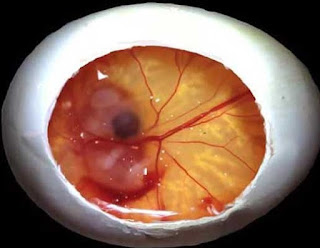
Improvement of Eye in Chick
The principal indication of the advancement of the eyes is a swelling at the parallel sides of the prosencephalon. These are the basics of the optic vesicles which lie underneath the head ectoderm. Then, the distal aspect of every optic vesicle (the future tactile layer) invaginates and presses against the proximal part (the future shade layer of the retina, iris and ciliary body).
This outcomes in the development of the optic cup, the end of the first lumen of the optic vesicle and the arrangement of another lumen, the future glassy chamber.
The focal point is shaped from the focal point placode, a thickening of the ectoderm framed because of an inductive sign from the optic cup. The focal point sinks underneath the outside of the ectoderm, the last turning into the cornea.
As the focal point keeps on developing, the cells in the thickened locale lose their capacity to partition and get changed over into filaments that will end up being the center of the grown-up focal point. New strands are framed from the cells at the fringe of the focal point which partition quickly and get masterminded in concentric circles around the first center.
When of incubating there are three concentric layers of strands, the center, the middle of the road layer of sporadically orchestrated filaments, and the outspread layers which keep on developing in the wake of bring forth. The focal point case, which is an extracellular material with a high collagenous segment, begins to frame about day 7. The ciliary body grows near the focal point, its job being to mystery


