Focal Nervous System
During undeveloped turn of events, the CNS initially shows up as a straightforward cylinder, the neural cylinder, which reaches out down the dorsal middle arrangement of the building up incipient organism’s body.
Cerebrum
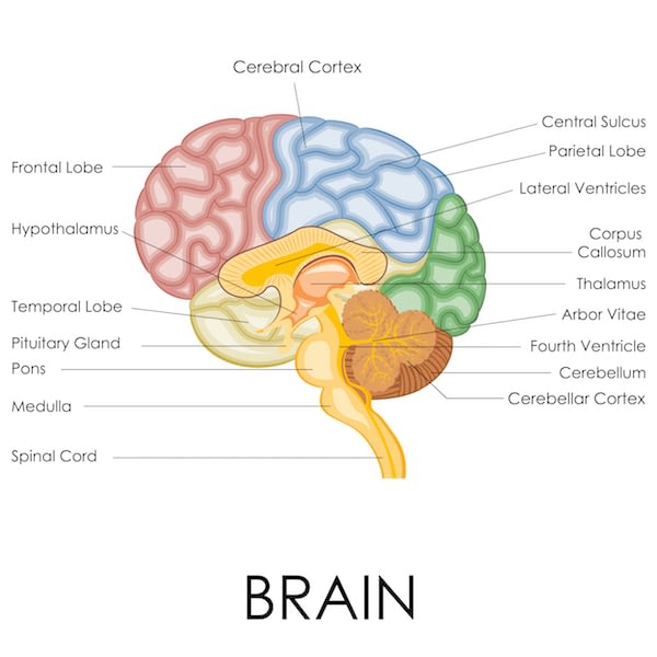
Since the cerebrum is the biggest and most complex mass of sensory tissue in the body, it is normally examined as far as its four significant locales – cerebral halves of the globe, diencephalon, mind stem, and cerebellum.
Cerebral Hemispheres
The matched cerebral halves of the globe, aggregately called cerebrum, are the most unrivaled piece of the mind, and together are significantly bigger than the other three cerebrum areas consolidated.
1.Gyri. The whole surface of the cerebral sides of the equator displays raised edges of tissue called gyri, isolated by shallow sections called sulci.
2.Fissures. Less various are the more profound sections of tissue called gaps, which separate huge areas of the mind; the cerebral halves of the globe are isolated by a solitary profound crevice, the longitudinal gap.
3.Lobes. Different gaps or sulci partition every half of the globe into various projections, named for the cranial bones that lie over them.
4.Regions of cerebral half of the globe. Each cerebral half of the globe has three fundamental districts: a shallow cortex of dark issue, an inner white issue, and the basal cores.
5.Cerebral cortex. Discourse, memory, legitimate and enthusiastic reaction, just as awareness, translation of sensation, and deliberate development are on the whole elements of neurons of the cerebral cortex.
6.Parietal flap. The essential physical tangible territory is situated in the parietal flap back to the focal sulcus; driving forces going from the body’s tactile receptors are limited and deciphered here.
7.Occipital flap. The visual region is situated in the back piece of the occipital flap.
8.Temporal flap. The hear-able region is in the fleeting flap circumscribing the horizontal sulcus, and the olfactory territory is discovered somewhere inside the transient projection.
9.Frontal projection. The essential engine zone, which permits us to intentionally move our skeletal muscles, is foremost to the focal sulcus in the front projection.
10. Pyramidal parcel. The axons of these engine neurons structure the significant deliberate engine plot the corticospinal or pyramidal parcel, which slips to the line.
11. Broca’s region. A particular cortical zone that is associated with our capacity to speak, Broca’s region, is found at the base of the precentral gyrus (the gyrus front to the focal sulcus).
12. Speech territory. The discourse region is situated at the intersection of the worldly, parietal, and occipital flaps; the discourse region permits one to sound out words.
13. Cerebral white issue. The more profound cerebral white issue is make out of fiber plots conveying motivations to, from, and inside the cortex.
14. Corpus callosum. One enormous fiber lot, the corpus callosum, associate the cerbral halves of the globe; such fiber lots are called commisures.
15. Fiber parcels. Affiliation fiber lots associate zones inside a half of the globe, and
projection fiber parcels interface the cerebrum with lower CNS focuses.
16. Basal cores. There are a few islands of dark issue, called the basal cores, or basal ganglia, covered profound inside the white matter of the cerebral halves of the globe; it controls the intentional engine exercises by adjusting directions shipped off the skeletal muscles by the essential engine cortex.
Diencephalon
The diencephalon, or interbrain, sits on the mind stem and is encased by the cerebral halves of the globe.
2.Hypothalamus. The nerve center makes up the floor of the diencephalon; it is a significant autonomic sensory system community since it assumes a part in the guideline of internal heat level, water equilibrium, and digestion; it is additionally the middle for some drives what’s more, feelings, and all things considered, it is a significant piece of the purported limbic framework or “enthusiastic instinctive mind”; the nerve center additionally directs the pituitary organ and produces two hormones of its own.
3.Mammillary bodies. The mammillary bodies, reflex focuses engaged with olfaction (the feeling of smell), swell from the floor of the nerve center back to the pituitary organ.
4.Epithalamus. The epithalamus structures the top of the third ventricle; significant pieces of the epithalamus are the pineal body (part of the endocrine framework) and the choroid plexus of the third ventricle, which shapes the cerebrospinal liquid.
Cerebrum Stem
The cerebrum stem is about the size of a thumb in distance across and roughly 3 inches in length.
1.Structures. Its structures are the midbrain, pons, and the medulla oblongata.
2.Midbrain. The midbrain reaches out from the mammillary bodies to the pons poorly; it is made out of two swelling fiber lots, the cerebral peduncles, which pass on sliding and climbing motivations.
3.Corpora quadrigemina. Dorsally found are four adjusted distensions called the corpora quadrigemina in light of the fact that they help some anatomist to remember two sets of twins; these protruding cores are reflex focuses engaged with vision and hearing.
4.Pons. The pons is an adjusted structure that distends just underneath the midbrain, and this zone of the mind stem is generally fiber lots; nonetheless, it has significant cores associated with the control of relaxing.
5.Medulla oblongata. The medulla oblongata is the most sub-par some portion of the cerebrum stem; it contains cores that manage crucial instinctive exercises; it contains focuses that control pulse, circulatory strain, breathing, gulping, and regurgitating among others.
6.Reticular development. Expanding the whole length of the mind stem is a diffuse mass of dark issue, the reticular development; the neurons of the reticular arrangement are associated with engine control of the instinctive organs; an exceptional gathering of reticular development neurons, the reticular enacting framework (RAS), assumes a function in awareness and the conscious/rest cycles.
CEREBELLUM
The huge, cauliflower-like cerebellum extends dorsally from under the occipital projection of the cerebrum.
1.Structure. Like the cerebrum. the cerebellum has two halves of the globe and a tangled surface; it likewise has an external cortex comprised of dim issue and an inward area of white issue.
2.Function. The cerebellum gives exact planning to skeletal muscle action and controls our equilibrium and balance.
3.Coverage. Strands arrive at the cerebellum from the harmony mechanical assembly of the internal ear, the eye, the proprioceptors of the skeletal muscles and ligaments, and numerous different regions.
Insurance of the Central Nervous System
Sensory tissue is delicate and sensitive, and the indispensable neurons are harmed by even the smallest weight, so nature has attempted to secure the cerebrum and the spinal line by walling them in inside bone (the skull and vertebral segment), films (the meninges), and a watery pad (cerebrospinal liquid).
MENINGES
The three connective tissue layers covering and ensuring the CNS structures are the meninges.
1.Dura mater. The furthest layer, the rugged dura mater, is a twofold layered film where it encompasses the cerebrum; one of its layer is connected to the inward surface of the skull, shaping the periosteum (periosteal layer); the other, called the meningeal layer, frames the peripheral covering of the mind and proceeds as the dura mater of the spinal rope.
2.Falx cerebri. In a few places, the internal dural film stretches out internal to frame a crease that appends the mind to the cranial pit, and one of these folds is the falx cerebri.
3.Tentorium cerebelli. The tentorium cereberi isolates the cerebellum from the cerebrum.
4.Arachnoid mater. The center layer is the weblike arachnoid mater; its threadlike augmentations length the subarachnoid space to append it to the deepest film.
5.Pia mater. The sensitive pia mater, the deepest meningeal layer, sticks firmly to the outside of the cerebrum and spinal rope, following each overlap.
CEREBROSPINAL FLUID
Cerebrospinal liquid (CSF) is a watery “stock” comparative in its cosmetics to blood plasma, from which it structures.
1.Contents. The CSF contains not so much protein but rather more nutrient C, and glucose.
2.Choroid plexus. CSF is consistently framed from blood by the choroid plexuses; choroid plexuses are bunches of vessels swinging from the “rooftop” in every one of the cerebrum’s ventricles.
3.Function. The CSF in and around the cerebrum and line shapes a watery pad that shields the delicate sensory tissue from blows and other injury.
4.Normal volume. CSF structures and depletes at a steady rate with the goal that its ordinary weight and volume (150 ml-about a large portion of a cup) are kept up.
5.Lumbar tap. The CSF test for testing is gotten by a technique called lumbar or spinal tap;because the withdrawal of liquid for testing diminishes CSF liquid weight, the patient must stay in a level position (resting) for 6 to 12 hours after the methodology to forestall a distressingly difficult “spinal migraine”.
THE BLOOD-BRAIN BARRIER
No other body organ is so totally subject to a steady inward climate just like the mind, thus the blood-cerebrum hindrance is there to secure it.
1.Function. The neurons are kept isolated from bloodborne substances by the purported blood-cerebrum hindrance, made out of the most un-porous vessels in the entire body.
2.Substances permitted. Of water-dissolvable substances, just water, glucose, and fundamental amino acids go effectively through the dividers of these vessels.
3.Prohibited substances. Metabolic squanders, for example, poisons, urea, proteins, and most medications are kept from entering the mind tissue.
4.Fat-dissolvable substances. The blood-mind boundary is for all intents and purposes pointless against fats, respiratory gases, and other fat-dissolvable particles that diffuse effectively through all plasma layers.


